
You need to have grit for animal husbandry. Over the years, my family and I have developed quite a passion for our goats. We continue to improve our herd, learn from our mentors, expand and grow our operations, and exhaustively research best practices.
Despite our best efforts, we still seem to encounter plenty of firsts, especially true now - during our biggest year yet in terms of growing and changing our herd.
Thankfully, we are still disease-free, as we have been very careful about our biosecurity. But we have certainly had our share of drama this year, as outlined in my previous post about calcium deficiency.
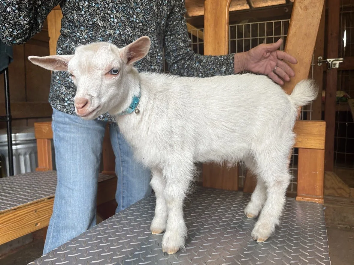
The purpose of this post is to talk about our latest case of umbilical hernia, which presented in our last buckling of the season.
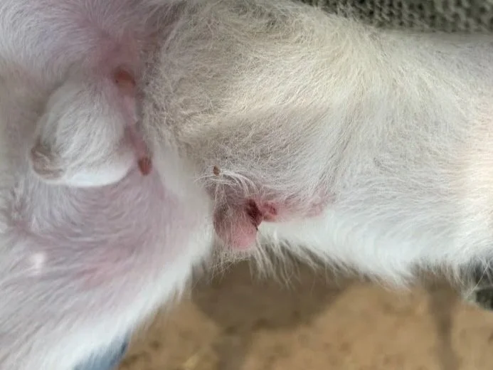
I noticed a small protrusion right next to his healing umbilical cord when he was about two weeks old. I already had payment for him as a buckling, so we were concerned on multiple levels. We decided to keep a close eye on it because some small hernias can close on their own as the kid grows. Unfortunately, that was not the case with this little guy.
He is currently five weeks old, and the hernia progressed over the weeks. While it is still relatively small, it is about four times the size it was when we first noticed it.
We knew a little bit about hernias from our prior research, but now it was time for a deep dive!
What I learned wasn't all that surprising, but it did require me to notify the buyer and offer her a refund. She thanked me for my honesty and accepted. Little Ghost will now become a wether rather than a breeding buck - more for the physical stress he would be under during rut than anything else. There may be a hereditary component to umbilical hernias - although research on this is very limited and inconclusive. As a precaution, we will not breed this doe to the same buck again, and we will keep a close eye on future offspring.
Because I don't ask you to take my word for it, I've put together some information below about the different types of hernias, their causes, symptoms, and treatment options. I've included veterinary references at the bottom for anyone who wants to dig a little deeper.
What Exactly Is a Hernia?
A hernia occurs when an internal organ, tissue, or portion of the intestine pushes through an opening or weakness in the surrounding muscle or connective tissue.
Most of us are already familiar with the term and it is no different than in humans. Think of it as a hole or weak spot in the abdominal wall that allows something inside the abdomen to push outward. Depending on the location and size of the opening, the hernia may be barely noticeable or quite large.
A hernia generally consists of three components:
The hernia ring: The opening or defect in the muscle wall.
The hernia sac: The tissue covering the protruding contents.
The hernia contents: What has pushed through the opening, which may include fat, connective tissue, or intestines.
This distinction is important because the size of the external bulge doesn't necessarily tell you how serious the condition is. A veterinarian needs to determine what is inside the hernia and whether it can be safely returned to the abdominal cavity.
Types of Hernias in Goats
There are several types of hernias that can occur in goats. Some are congenital (present at birth), while others develop later because of trauma, injury, infection, or weaknesses in the abdominal wall.
1. Umbilical Hernia
This is the one we are dealing with!
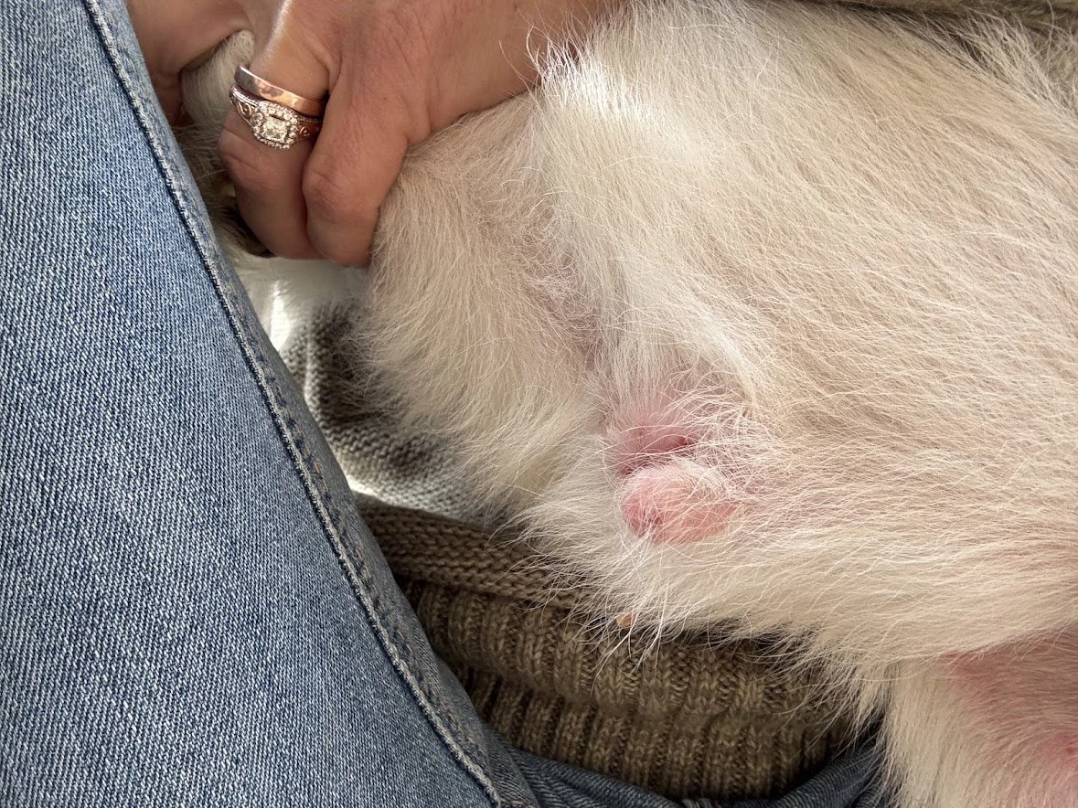
An umbilical hernia occurs when the opening in the abdominal wall where the umbilical cord passed during pregnancy does not close properly after birth. As a result, abdominal contents can protrude through the opening near the navel.
Umbilical hernias can be congenital or develop secondary to infection of the umbilical structures. A genetic predisposition is suspected in some livestock species, although the evidence for a hereditary cause specifically in goats is not well established.
What to look for:
A soft lump or bulge near the belly button.
A swelling that may become more noticeable as the kid grows.
A bulge that may change in size when the kid stands, lies down, or strains.
Occasionally, pain, warmth, or tenderness if complications develop.
Small hernias may close naturally during early development. However, persistent or enlarging hernias need veterinary evaluation. An opening that traps intestinal tissue can become an emergency.
Treatment:
Treatment depends on the size of the hernia ring, what is protruding through it, and whether the contents can be returned safely.
A veterinarian may recommend monitoring a very small hernia, but larger or persistent hernias generally require surgical correction. In some cases, veterinarians may use other closure techniques, but these are not appropriate for every animal.
Our little buckling has been to the veterinarian who confirmed the hernia but also noted that the protrusion is hard rather than soft, indicating mesenteric fat is all that has pushed through. He palpatated the protrusion and didn’t sense anything else, and the goat showed no idication of pain, so he said this fat is like a belly bandaid. He recommended monitoring.
Surgery on ruminants is highly risky, and when it comes to livestock, you will spend more money on operating than the goat is worth. He should be just fine, and if it gets worse we still have the option to operate, but is is never recommeded to start with surgery.
2. Inguinal Hernia
An inguinal hernia occurs when abdominal contents push through the inguinal canal, which is located in the groin area.
This type of hernia is particularly concerning in male animals because the inguinal canal is associated with the passage of the spermatic cord.
What to look for:
Swelling in the groin area.
An unusual bulge near the upper thigh or genital region.
Discomfort or pain.
In some cases, swelling that extends into the scrotum.
Treatment:
Surgical correction may be necessary, depending on the severity and contents of the hernia. If intestinal tissue becomes trapped, emergency intervention may be required.
3. Scrotal Hernia
A scrotal hernia is essentially an extension of an inguinal hernia in which abdominal contents, often a portion of the intestine, descend into the scrotum.
This can be particularly problematic in breeding males. Besides the possibility of intestinal entrapment, the abnormal positioning of abdominal contents can interfere with normal testicular function.
What to look for:
An unusually enlarged scrotum.
Swelling that may be more pronounced on one side.
A soft or fluid-like swelling.
Discomfort, particularly if the intestine becomes trapped.
Treatment:
Veterinary examination is essential. Surgical correction may be considered, but the animal's future breeding potential must also be evaluated. Castration may be recommended depending on the condition and the surgical approach.
4. Ventral or Abdominal Hernia
Unlike an umbilical hernia, which occurs specifically at the navel, a ventral or abdominal hernia can occur anywhere along the abdominal wall.
These are often associated with trauma, such as being kicked, falling, or experiencing an injury that damages the abdominal muscles.
What to look for:
A noticeable bulge along the belly or side.
Swelling that may become larger over time.
A soft area where the abdominal wall should normally feel firm.
Pain or discomfort, depending on the severity.
Treatment:
Treatment depends on the size and location of the defect. Some smaller hernias may be monitored, while larger defects generally require surgical repair.
5. Diaphragmatic Hernia
This is a much less common and more serious type of hernia.
The diaphragm is the muscle separating the chest cavity from the abdominal cavity. A diaphragmatic hernia occurs when abdominal organs move through an opening in the diaphragm and into the chest cavity.
These can be congenital or acquired following significant trauma.
What to look for:
Difficulty breathing.
Rapid or labored breathing.
Poor growth or failure to thrive.
Exercise intolerance.
General signs of distress.
Treatment:
This condition requires veterinary evaluation and may require surgical repair. The prognosis depends on the organs involved, the severity of the defect, and the animal's overall condition.
How Are Hernias Diagnosed?
Although some hernias are fairly obvious, it is important not to assume every lump around a kid's belly is a hernia.
Your vetrinarian may perform a physical examination including:
Palpation: Feeling the swelling to determine its size, firmness, tenderness, and whether the contents can be gently returned through the opening.
Checking the hernia ring: Determining the size of the actual opening in the abdominal wall.
Ultrasound: Helping identify what is inside the hernia, including whether intestinal loops or other abdominal structures are involved.
Checking for infection: An infected umbilicus can cause swelling that resembles a hernia. These conditions can also occur together.
This last point is especially important. Navel infections, sometimes called navel ill, can involve the umbilical structures and potentially contribute to the development of a hernia.
When Is a Hernia an Emergency?
A hernia becomes particularly dangerous when the contents become trapped. This is called an incarcerated hernia. If blood supply to the trapped tissue is compromised, it can become a strangulated hernia.
When this happens, the affected tissue can begin to die, and the condition can quickly become life-threatening.
Watch for these warning signs:
Sudden enlargement of the hernia.
A firm, hot, or painful swelling.
Loss of appetite.
Abdominal discomfort or bloating.
Teeth grinding.
Depression or lethargy.
Straining or difficulty passing manure.
Signs of severe distress.
If you suspect a trapped or strangulated hernia, contact your veterinarian immediately. Do not attempt to force the contents back into the abdomen yourself.
Even if a hernia appears relatively harmless, it is worth having it evaluated early. Waiting until it becomes an emergency can make treatment much more complicated.
Can Hernias Be Prevented?
Unfortunately, not all hernias can be prevented, particularly those involving congenital defects.
However, there are some things we can do as goat owners to reduce the risk of complications:
Maintain clean kidding areas. A clean, dry environment helps reduce the risk of umbilical infections.
Properly disinfect newborn navels. Follow appropriate veterinary recommendations for umbilical care immediately after birth.
Monitor newborns. Make checking the navel part of your regular newborn kid examination.
Watch for swelling. Don't assume every bump is normal or that it will disappear on its own.
Keep breeding records. If a congenital defect appears, document the dam, sire, and any affected offspring.
Make responsible breeding decisions. Avoid breeding animals with known congenital defects until you have discussed the potential hereditary implications with your veterinarian.
One thing I learned during this research is that we shouldn't automatically assume an umbilical hernia is genetic in goats. There is evidence of hereditary involvement in some livestock species, but the research specifically identifying a genetic predisposition in goats is limited. That doesn't mean we should ignore the possibility. It simply means we need to be careful about drawing conclusions from one case.
To wrap it up…
This is another one of those experiences that reminds me why raising livestock requires constant learning. You can read all the books, follow all the recommendations, consult your mentors, and do everything you believe is right, but occasionally something unexpected happens.
And when it does, you learn.
I hope this information helps another goat owner recognize an umbilical hernia early and understand what to look for. If you notice an unusual swelling on one of your kids, don't just wait and hope it goes away. Have your veterinarian take a look.
We'll keep you posted on our little guy's surgery and recovery!
References and Further Reading
These are the veterinary and scientific resources I used when researching this topic:
1. Merck Veterinary Manual – Hernias in Animals. An overview of different hernia types, their anatomy, causes, and treatment considerations.
2. Smith, M.C. and Sherman, D.M. – Goat Medicine, 2nd Edition. A comprehensive veterinary reference covering goat health, diseases, and surgical conditions.
3. Diseases of the Gastrointestinal System – Veterinary Reference Chapter. Includes information on umbilical hernias, diagnosis, ultrasound examination, and surgical indications in ruminants.
4. Al-Sobayil, F.A. and Ahmed, A.F. (2007). Surgical Treatment for Different Forms of Hernias in Sheep and Goats. Journal of Veterinary Science, 8(2), 185–191. A clinical study examining surgical treatment and outcomes in sheep and goats.
5. Merck Veterinary Manual – Breeding and Parturition of Goats. Includes information on newborn kid care and preventing umbilical infections.